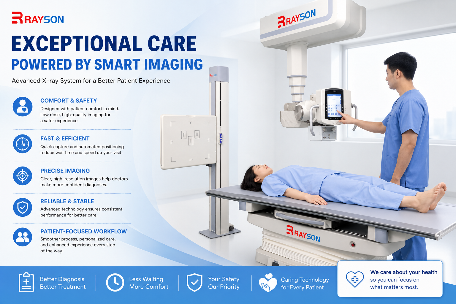
Unlike conventional column-mounted DR systems, the X-ray tube of the suspension dynamic DR is mounted on an overhead rail, allowing both the X-ray tube and detector to move over a large range in three-dimensional space. This enables flexible adjustment of projection angles and irradiation height, easily covering all body parts—particularly special angles that are difficult to achieve with traditional equipment, such as open-mouth views of the cervical spine and lateral views of the sacrococcyx.
For patients with severe trauma, intense pain, postoperative mobility restrictions, or special body types (e.g., obesity), who may struggle to cooperate with standard positions, the suspension dynamic DR can freely adjust the X-ray tube angle and height. This minimizes patient discomfort and the risk of secondary injury, ensuring critical images are obtained while the patient is in a relatively comfortable or stable posture.
Additionally, due to positional limitations, traditional equipment often requires repeated adjustments and multiple exposures, inadvertently increasing patient radiation dose. In contrast, the suspension dynamic DR, with its flexible positioning capabilities, can quickly complete precise irradiation. This not only significantly improves examination efficiency but also reduces patient radiation dose.
Static radiography can only capture a single instant image, making many hidden fractures and joint instability difficult to detect. Suspended dynamic DR features real-time dynamic imaging, allowing physicians to dynamically observe bone movement, capture subtle lesions during motion, and obtain high-definition spot images of critical moments in real time. In emergency trauma screening, it can efficiently detect easily overlooked hidden fractures. For conditions such as rotator cuff injuries, ankle joint instability, and lumbar spine dynamic instability, it intuitively displays abnormal changes in bone and joint movement, providing key evidence for functional diagnosis.
A single suspended dynamic DR system can cover the majority of orthopedic imaging examination scenarios, meeting the diagnostic and therapeutic needs of multiple departments such as emergency trauma orthopedics, spinal surgery, joint surgery, sports medicine, and rehabilitation orthopedics. It can quickly complete emergency screening for limb fractures, pelvic injuries, and occult fractures. It can accurately evaluate spinal diseases such as scoliosis, cervical spondylosis, and lumbar lesions. At the same time, it can efficiently perform multi-joint functional position imaging and stability assessment for knees, hips, ankles, shoulders, etc. It is also suitable for follow-up examinations such as postoperative rehabilitation in orthopedics and dynamic monitoring of joint function.
In addition, the device features panoramic stitching functionality, which allows for segmented radiography of the spine and lower limbs followed by stitching to fully present the overall morphology of the entire spine or lower limbs. This provides high-precision imaging support for adolescent scoliosis screening. This integrated application not only reduces the hospital's multi-device procurement costs and machine room space occupation but also significantly optimizes the department's examination workflow.
With ongoing intensification of orthopedic competition across hospitals at all levels, basic radiography alone can no longer meet the requirements for high-quality diagnosis and treatment. Suspended dynamic DR addresses the technical gap in traditional orthopedic imaging by enabling dynamic functional assessment, helping hospitals expand characteristic treatments projects such as dynamic joint evaluation. Whether it is the orthopedic department of a comprehensive hospital, a rehabilitation hospital, or an orthopedic specialty hospital, this equipment can be relied upon to improve the diagnostic and treatment chain, enhance the precision of specialty diagnosis and treatment, build differentiated technical advantages, and support the upgrading of orthopedic specialty capabilities within the hospital.
As orthopedic imaging diagnosis and treatment continues to move toward greater precision and efficiency, suspended dynamic DR empowers clinical practice with multiple unique advantages, continuously safeguarding precise orthopedic diagnosis and supporting the high-quality development of medical institutions.